

❋
Ureteral Strictures
What the ureter is, and what a stricture does
The ureters are the two narrow tubes — each about the width of a drinking straw — that carry urine from your kidneys down to your bladder.
A ureteral stricture is a narrowed segment of one of those tubes, caused by scar tissue. When it narrows, urine backs up behind it. The kidney above swells, a condition called hydronephrosis, and over time that back-pressure damages the kidney.
Why this one is dangerous quietly
Some men and women with a ureteral stricture have flank pain, infections, or blood in the urine. Many have no symptoms at all.
A kidney can lose most of its function from a slow blockage without ever hurting. It is not unusual for a stricture to be found incidentally on a scan done for something else, at a point where kidney function has already been lost permanently.
This is why we take an incidentally discovered stricture seriously even when you feel completely well, and why we measure how well each kidney is actually working before deciding what to do.
What causes them
Kidney stones, especially a stone that sat impacted in one place, and injury from the ureteroscopy used to treat it. This is now one of the most common causes.
Previous surgery in the abdomen or pelvis — gynecologic, colorectal, vascular, or urologic. The ureter runs close to a great deal of important anatomy.
Radiation to the pelvis or abdomen.
Endometriosis, which can encase and constrict the ureter.
Retroperitoneal fibrosis, an inflammatory process that traps the ureters.
Kidney transplant, where the join between the transplanted ureter and the bladder can narrow.
Loss of blood supply to a segment of ureter, which is often what underlies the scarring.
No identifiable cause, in a minority of cases.
How we evaluate it
We need three things before recommending a repair: where the narrowing is, how long it is, and how much function the kidney behind it still has.
CT urogram — a CT scan with contrast that outlines the entire collecting system.
A renal scan (MAG3 or similar) — measures how much each kidney contributes and whether the blockage is significant. If a kidney has lost nearly all function, reconstruction may not be worthwhile and other options make more sense.
Retrograde or antegrade pyelogram — contrast injected from below or above to map the narrowed segment precisely.
Ureteroscopy — a small camera passed up the ureter to see the segment directly, and to take biopsies if there is any concern about a tumor rather than scar.

Living with a stent — and why it isn't a treatment
Most people reach a reconstructive urologist already carrying a ureteral stent or a nephrostomy tube.
Both are excellent temporary solutions. A stent keeps urine flowing while we plan. A nephrostomy tube drains the kidney directly through the back when a stent won't pass.
But neither fixes anything, and both have costs. Stents commonly cause flank pain when you urinate, urinary urgency, blood in the urine, and infections. They must be exchanged every few months, indefinitely, each time under anesthesia. Nephrostomy tubes mean an external bag and regular exchanges.
Many people are told this is simply how it will be from now on. For most people, it isn't. An indefinite cycle of stent exchanges is a holding pattern, not an endpoint, and it is worth getting an opinion from a surgeon who reconstructs ureters before accepting it as permanent.
Your treatment options
Endoscopic treatment
Balloon dilation or endoureterotomy — stretching or incising the narrowed segment from the inside — can be done through a scope, without incisions.
These work best for short, recently formed strictures with good blood supply, and they are reasonable to try in the right situation. The limitation is the same as with any scar: stretching it doesn't remove it. Long-term durability is modest, and results are poor for longer strictures, strictures related to radiation, and strictures where the blood supply is compromised.
Robotic reconstruction
This is what we do, and it is the part of ureteral stricture care that is least widely available.
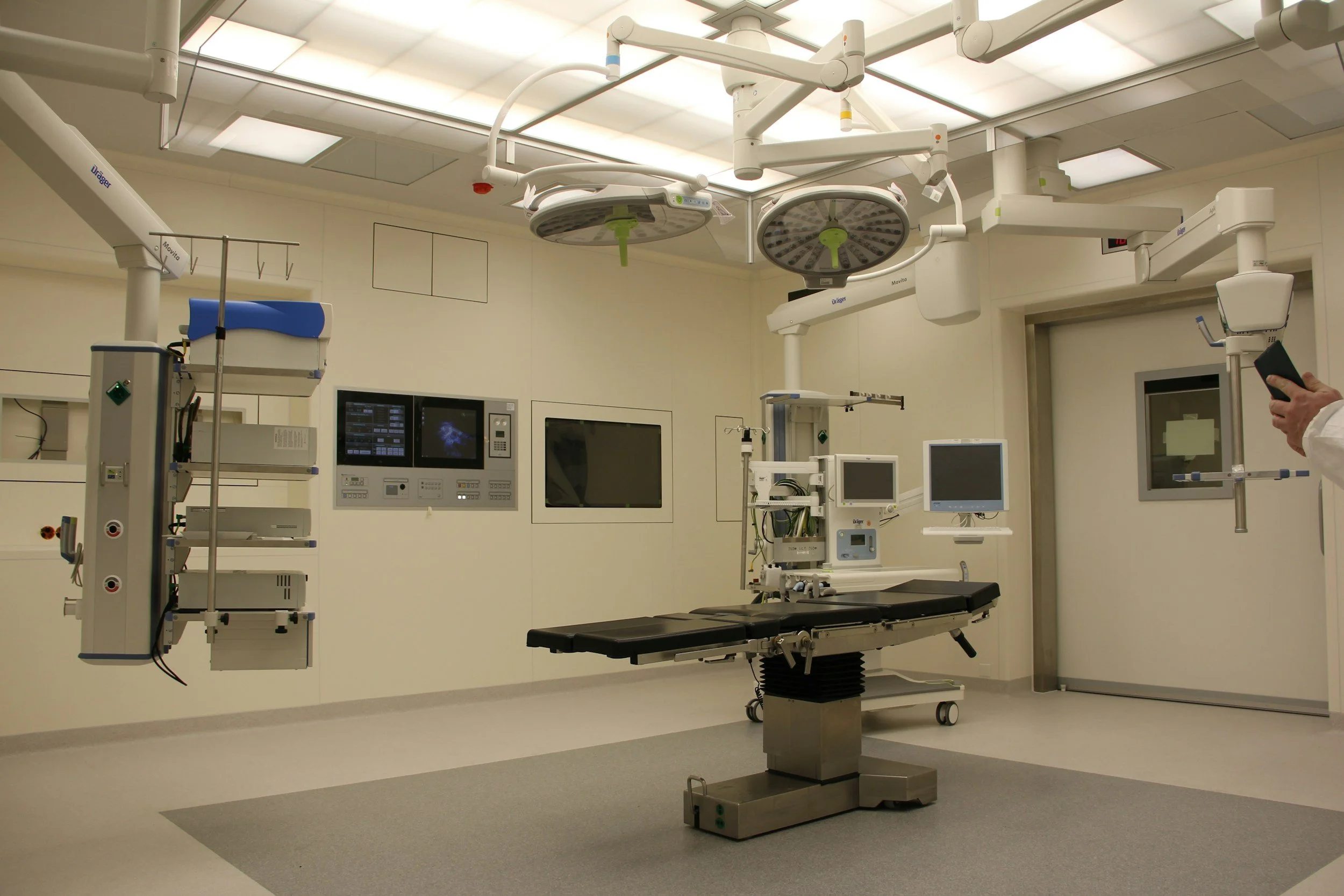
Reconstructing a ureter used to mean a large open incision, several days in the hospital, and a long recovery. Robotic surgery has changed that substantially: the same operations are now done through several small incisions, usually with a one- or two-night stay.
Why the robot matters here specifically
The ureter is small, deep, and delicate, and its blood supply is fragile. Robotic surgery provides high magnification and instruments that articulate like a wrist, which makes precise suturing of a narrow tube in a deep space far more feasible than through a long open incision.
It also allows the use of indocyanine green fluorescence — a dye given during surgery that makes well-perfused tissue glow under a special camera. This lets us see, in real time, where the healthy blood supply actually ends. Since poor blood supply is what causes strictures to recur, being able to see it rather than guess at it directly improves the odds that the repair lasts.
The operations
Which one you need depends entirely on where the stricture is and how long it is.
Ureteroureterostomy. For short strictures. The scarred segment is cut out and the two healthy ends are sewn back together.
Ureteral reimplantation. For strictures near the bladder. The healthy ureter is detached and re-implanted into the bladder. If it doesn't reach comfortably, the bladder is mobilized up toward it — a psoas hitch — or a flap of bladder wall is tubularized to bridge the gap — a Boari flap.
Buccal mucosa graft ureteroplasty. For long strictures in the upper or middle ureter that are too long to simply cut out. The narrowed segment is opened lengthwise and widened with a patch of tissue taken from the inside of your cheek, then wrapped in omentum — the body's own fatty apron — to give the graft a rich blood supply.
This is the same principle used to rebuild the urethra, applied to the ureter. It is a relatively new operation, performed at a limited number of centers, and it has meaningfully changed what's possible: strictures that once meant replacing the ureter with bowel can now often be repaired with the patient's own tissue. Multi-institutional results have been strong, with the large majority of patients free from further intervention at intermediate-term follow-up.
Appendiceal onlay or interposition. On the right side, the appendix can be opened and used as a patch, or used as a tube to bridge a longer gap. It has its own blood supply, which is an advantage.
Ileal ureter. For very long or multiple strictures, a segment of small intestine can be reconfigured to replace the ureter entirely. This is a bigger operation with more long-term metabolic considerations, but it preserves kidneys that otherwise couldn't be saved.
Downward nephropexy — repositioning the kidney slightly lower — can add length when a repair is close to reaching.
We will tell you which of these we expect to do, and we will also tell you honestly that the final decision is sometimes made at the operating table once we can see the tissue.
Recovery
Typically one to two nights in the hospital
A ureteral stent stays in for roughly four to six weeks afterward, and is removed in the office
Most people are back to light activity within a week or two, and to full activity by about six weeks
If a cheek graft was used, your mouth will be sore for about a week
Imaging a few weeks after stent removal confirms the repair is draining, and we follow you with periodic imaging or a renal scan after that
Risks
Bleeding, infection, urine leak from the repair while it heals, injury to nearby structures, and recurrence of the stricture. Occasionally a repair planned as robotic needs to be converted to an open incision. If a graft is used, the mouth donor site is sore temporarily.
Common questions
Can my kidney recover? Often partly, depending on how long it's been obstructed and how much function remains. The renal scan gives us the honest answer before we operate.
Can I get rid of my stent for good? That is the entire point of reconstruction. Most people who have a successful repair are stent-free permanently.
What if my kidney is already mostly dead? Then reconstruction may not help you, and the conversation shifts to whether the kidney needs to come out or can simply be observed. We'd rather tell you that than perform an operation that won't change anything.
Is robotic surgery as good as open surgery? For ureteral reconstruction, published results are comparable, with less blood loss and shorter hospital stays. The important variable is how often the surgeon does the operation, not the platform.
I was told nothing more could be done. Get a second opinion from a reconstructive urologist. Complex ureteral reconstruction is a narrow subspecialty, and what's routine at a reconstructive center may not be offered elsewhere.
Questions worth asking at your consultation
How long is my stricture, and where exactly is it?
How much function does that kidney still have?
Which repair would you plan, and what's the backup if the tissue looks different than expected?
Would you use a graft, and where from?
How long will I have a stent afterward?
How many robotic ureteral reconstructions do you perform each year?
To schedule a consultation and discuss which option is right for you, please visit our CONTACT page.